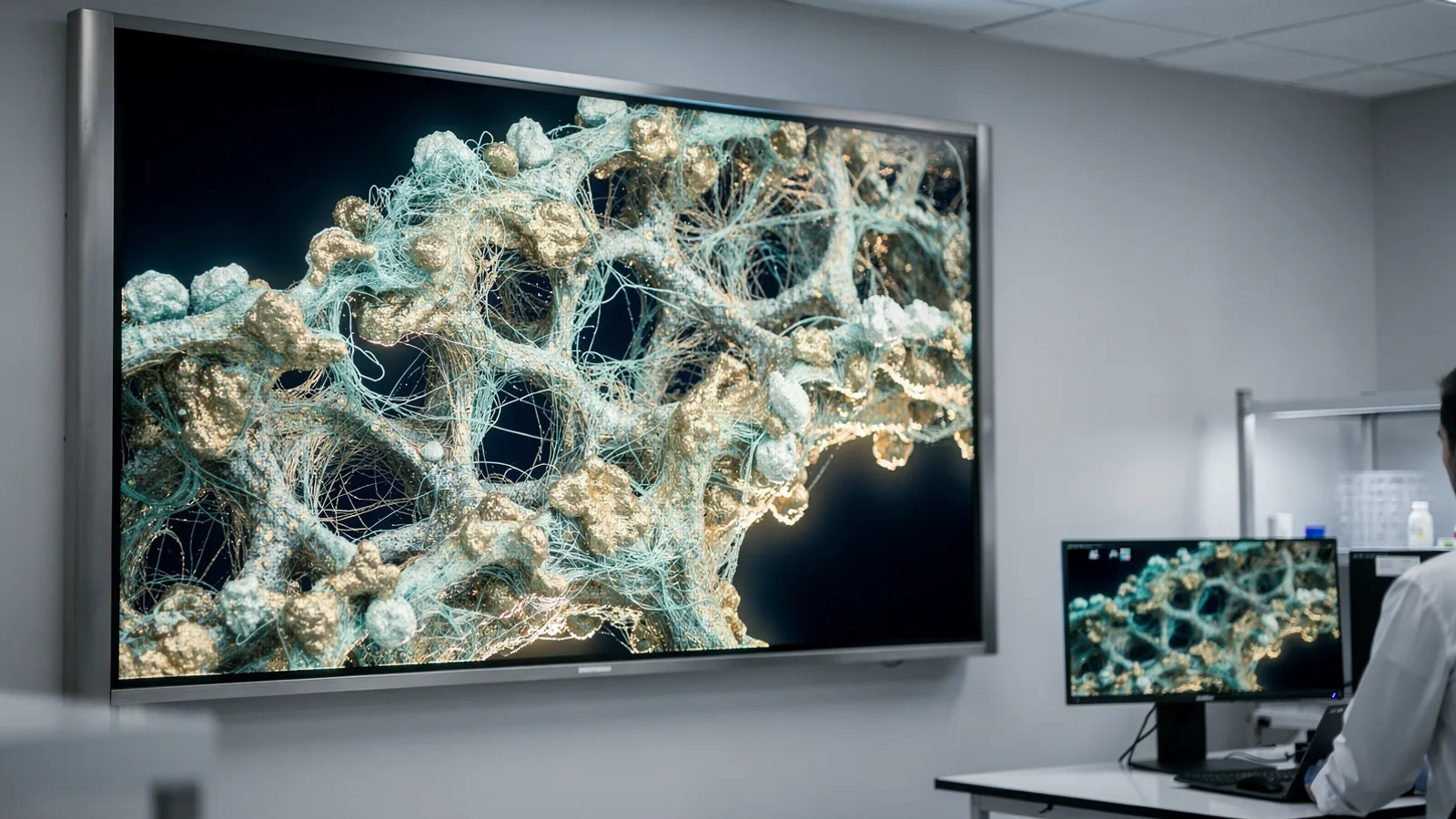
A Sharper Microscope Lets Texas A&M Scientists See Deep and Small at Once
BulletinTexas A&M scientists designed an imaging approach that captures nanoscale cellular structures deep within living biological tissue.
- 75 nmlateral resolution achieved by the method
- 5 timessharper than conventional two-photon microscopy
- 2024year of earlier foundational study
Texas A&M researchers have developed a microscopy technique capable of resolving fine cellular details deep inside living tissue at the same time, according to a report published in the Journal of Biomedical Optics.
COLLEGE STATION, Texas (Stratosyn) · The new technique resolves structural features smaller than 100 nanometers across, a scale roughly one thousand times thinner than a human hair. By maintaining clarity deep inside intact samples, the system preserves image quality without losing penetration depth.
The work was led by Aart Verhoef, an assistant professor of biophotonics in the Department of Soil and Crop Sciences within the College of Agriculture and Life Sciences. Verhoef, who also serves as a joint professor in the Department of Physics and Astronomy, completed the research with colleague Alma Fernandez Gonzalez.
New Optical Framework
The team published its findings in the Journal of Biomedical Optics after assembling the specialized configuration. Known formally as 2PE-ISM-SOFI, the system merges two-photon excited image scanning microscopy with super-resolution optical fluctuation imaging.
Rather than building unfamiliar hardware from scratch, the researchers layered these two established techniques directly onto a standard two-photon microscope. This design provides a practical way to boost laboratory performance using familiar optical instruments.
Sharper Resolution
Under this configuration, the microscope delivers a lateral resolution of about 75 nanometers inside living biological specimens. That degree of visual precision makes the output roughly five times sharper than what conventional two-photon microscopy normally achieves.
The technical foundation for this achievement stems from earlier groundwork documented in a 2024 study published in PRX Life. The new progression demonstrates that extreme detail and substantial imaging depth can be attained simultaneously within whole biological samples.
Applications in Agriculture
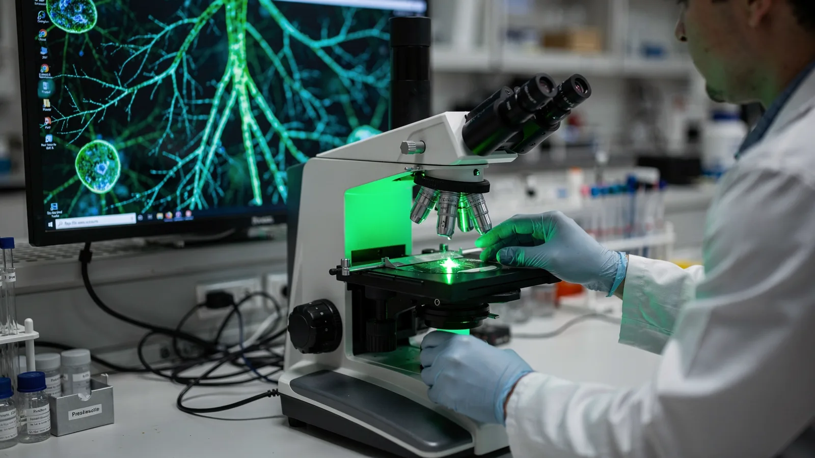
The advanced imaging method holds direct potential for expanded research across plant and soil biology. Scientists can apply the sharper views to observe intricate root structures in living tissue.
The refined visibility also creates new opportunities to examine plant-microbe interactions at the cellular level. By offering clear views into living systems, the approach aids ongoing agricultural studies.
This bulletin is based on reporting and materials from Texas A&M Stories, rewritten in our own words.